FDG-PET Segmentation: Automated TMTV Calculation
Standardizing Total Metabolic Tumor Volume extraction to enhance predictive accuracy in oncology clinical trials.
Tumor Burden, a new imaging biomarker ready for decisional trials
"At Pixilib we see medical imaging as a leverage to enhance patients' healthcare and provide more accurate treatments."
Last year, PET/CT segmentation with Total Metabolic Tumor Volume (TMTV) calculations showed a strong prognosis value in various tumors. With TMTV, tumor burden can be quantified to evaluate a patient's outcomes.
For instance in Lymphomas, this biomarker is now being integrated into new patient risk stratification to build more tailored therapies — reducing toxicity for low-risk patients and increasing treatment efficacy for high-risk patients.
Our technology makes TMTV calculation accessible in prospective decisional trials to have real-time, centralized, expert-validated TMTV calculation that can be integrated for treatment adaptation.
Precision Algorithms for TMTV Extraction
Our image processing pipeline is optimized to handle the challenges of FDG PET across multicenter studies, ensuring that your Total Metabolic Tumor Volume data is reproducible, standardized and medically validated (human in the loop).
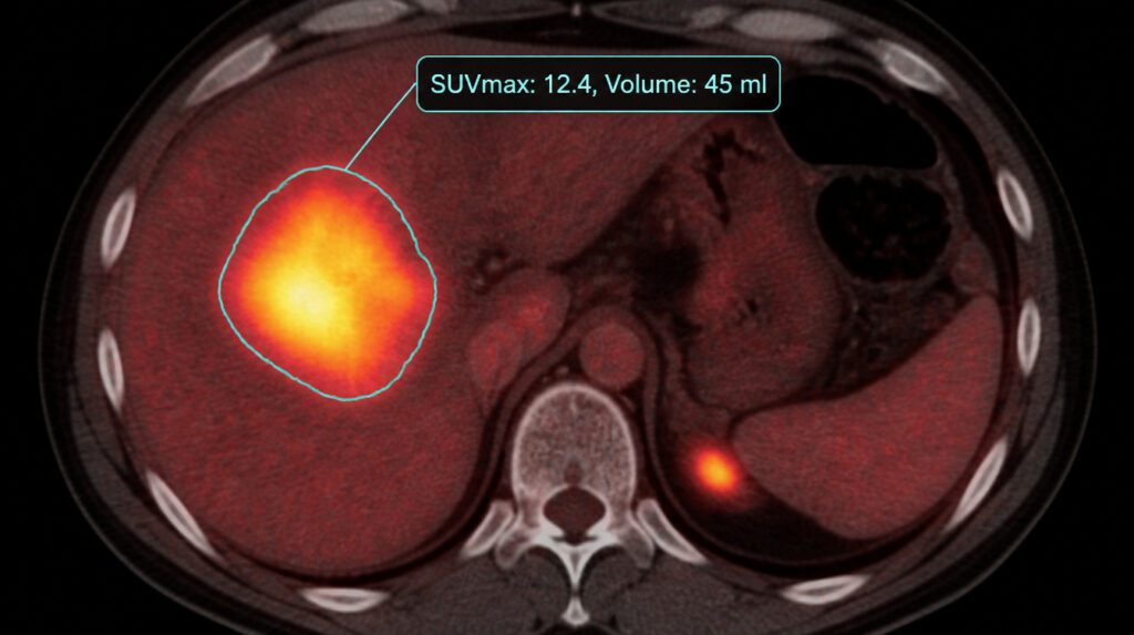
Automated Lesion Detection & Segmentation
Pixilib's algorithms identify hypermetabolic foci while filtering out physiological uptake (e.g., brain, heart, bladder). This automated Tumor Segmentation ensures:
- Consistency eliminating the inter-reader variability inherent in manual contouring
- Speed calculating the Total Metabolic Tumor Volume in seconds, even for patients with high metastatic dissemination
- Validity and Accuracy AI delineates lesions, then validated thresholding methods (e.g., 41% SUVmax or SUV 2.5) are applied according to the study protocol
Human In The Loop, Tumor Burden Assessment
Our AI algorithm is integrated with a real DICOM viewer, making it possible for physicians to review AI results and edit them — remove false positives, add missing lesions.
At Pixilib we don't think there will be a perfect AI capable of 100% accuracy in segmentation tasks. We believe in complementarity: AI algorithms removing the unneeded work burden of standard segmentation tasks, and expert review focusing on more complicated cases and unusual uptake to deliver a final medically validated TMTV.
Our algorithms are designed to reach this complementarity — building the best of AI and medical expert union.
Radiomics calculation pipeline
Once the tumor mass is segmented, TMTV is an important factor to extract — but not the only one. From this segmentation we extract many new biomarkers that could be integrated into clinical trials:
- Total Lesion Glycolysis (TLG)
- Tumor Spread (Dmax)
- Uptake intensity (SUVmax, SUVpeak, SUVmean)
- Shape features Surface, Surface-to-volume ratio, Elongation, Flatness, Compactness...
- Textural features
In combination with our CT segmentation algorithm, we also integrate differentiation of involved organs to calculate separate organ involvement burden and extract anthropomorphic quantifications.
Why Automate your FDG PET Analysis with GaelO?
From multicenter clinical trial imaging harmonization to ready-to-export TMTV metrics — three reasons to bring AI-validated tumor burden into your next protocol.
Predictive Power
TMTV and derived metrics are increasingly recognized as major surrogate endpoints for progression-free survival (PFS) and Overall Survival (OS).
Multicenter Harmonization
Our algorithms account for differences in PET/CT scanner sensitivity and reconstruction methods, delivering reproducible TMTV values regardless of the acquisition site.
Ready for Clinical Reports
Export your TMTV and derived metrics directly into your Electronic Data Capture (EDC) system or clinical study report — no manual transcription, no rounding errors.
A unified infrastructure for medical imaging research
From clinical trial centralization to research PACS and AI segmentation — three complementary products covering the full lifecycle of medical imaging in clinical research.
GaelO
Collection and centralized reading of clinical trials. From DICOM upload to Blind Independent Central Review with disease-specific criteria — GDPR/HIPAA compliant, fully auditable.
Discover GaelOGaelO Flow
Research PACS for retrospective and Real World Evidence studies. Orchestrate medical imaging data: query/retrieve, batch de-identification, AI inference pipelines and seamless export.
Discover GaelO Flow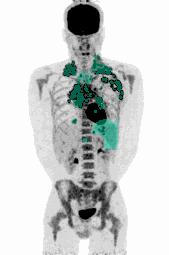
AI Segmentation Algorithms
FDG, PSMA, DOTATOC. Automated PET segmentation to extract Total Metabolic Tumor Volume (TMTV) human-in-the-loop, 100% medically validated, regulatory-ready.
Explore algorithms